We are moving!

After a few years being hosted on wordpress.com I feel the time has come when the site has outgrown the capabilities of the standard wordpress hosting. WordPress however also offers self-hosting, which gives many more benefits than the standard capabilities of wordpress.com.

So, over the next few days the site will be moving to http://vetsci.co.uk (live now!) – all the original content available here will be moved to the new site along with all comments etc. When the move is entirely complete this domain (vetsci.wordpress.com) will point to the new address (vetsci.co.uk) meaning all bookmarks, links, etc. will continue to function.

With the move comes a new design, as well as multiple new features for you!

If you have any thoughts on the new design, feel free to comment or get in touch – we’d love to hear from you, as for now you can continue to use this site or go and have a look at the new site over at http://vetsci.co.uk

Thank you for you continued support!

Surviving Sub-Zero

Introduction

Pure water has both a freezing point and melting point of 0˚C; however water also has colligative properties meaning that the freezing point can vary depending on the number of molecules dissolved in it. As the concentration of molecules increases, the freezing point decreases (and the boiling point increases). For example, pure water will have a higher freezing point than salt water. This is because the presence of dissolved molecules in the water makes the formation of hydrogen bonds less energetically favourable, such as those formed by ice crystals. The change in freezing point is known as freezing point depression.

Freezing Point Depression

The decrease in the temperature at which a fluid freezes (or freeze point depression) can be represented by ΔFp. To work out how ΔFp varies the equation below is used:

ΔFp= -1.86˚C x Osm

Where:

  • ΔFp = The change in freezing point
  • -1.86˚C = Constant
  • Osm = The osmolarity of the fluid

A 1M solution of glucose has an osmolarity of 1 Osm, a 0.5M solution of NaCl also has an osmolarity of 1 Osm (0.5M of Na and 0.5M of Cl). If we consider that seawater has contains 0.5M of NaCl, we can determine the ΔFp of seawater. This would give:

ΔFp= -1.86˚C x 1

ΔFp= -1.86˚C

Therefore the freezing point of seawater is -1.86˚C, which is lower than pure water due to the dissolved NaCl molecules.

Ice Nucleation

The formation of ice crystals usually requires a form of ‘catalyst’ known as an ice nucleating agent (INA). The presence of an INA allows water molecules to begin forming an ice crystal around it. Such agents include; dust, bacteria, proteins or already present ice crystals. Without the presence of an INA, much lower temperatures can be reached before freezing occurs, this is known as supercooling.

Supercooling

Supercooling is a process where liquids can reach very low temperatures before rapid freezing occurs, this is because there are no ice nucleating agents (INAs) present. Without an INA present, water molecules struggle to begin forming ice crystal and hence the fluid stays in liquid form.

For example, a beaker of non-distilled water is cooled slowly and freezes at around 0˚C. Another beaker of pure water is cooled and care is taken to remove all INAs. This requires using pure H20 and a clean, sterilised glass free from dust and impurities etc. The experiment must also be carried out in a clean atmosphere or the beaker must be covered to prevent INAs such as dust entering. If no INAs are present, then the beaker of water can be cooled past 0˚C, it is possible to reach -40˚C before spontaneous rapid freezing occurs. The point at which a liquid will freeze in the absence of INAs is known as its super cooling point.

Even though the 2nd beaker of water froze at -40˚C, its freezing point is still 0˚C, this means that spontaneous freezing can occur anywhere between 0 to -40˚C. Freezing post-freeze point can often be induced by disturbing the liquid i.e. tapping the glass.

Freeze Injuries

Freezing of internal fluids is dangerous and can cause damage to living organisms by a number of means:

  • Physical damage, for example, the formation of ice crystals which could pierce and burst the lining of small capillaries.
  • Freezing can lower the rate of blood flow (ischemia), which would reduce the rate at which tissues receive oxygen and nutrients. This in turn could lead to anoxia.
  • The thawing process can lead to the release of reactive oxygen species which can damage DNA
  • Freezing may also cause osmotic shock, this can lead to collapse of the cell membrane, swelling or bursting of the cell.

Osmotic Shock

Organisms without adaptations to cope in freezing conditions may experience osmotic shock, a cell damaging process caused by the formation of ice crystals in tissues. Typically concentration gradients between intracellular and extracellular fluids are at equilibrium; however at freezing temperatures the formation of ice crystals can alter this:

  • At temperatures below zero, ice crystals can form from the water in the extracellular fluids
  • The formation of the ice crystal reduces dissolved H20 and thus increases the concentration of the extracellular fluid
  • H20 within the intracellular fluids (i.e. the fluid within cells) leaves the cell along the concentration gradient to form equilibrium again, as a result the cell shrinks
  • As the ice crystal continues to grow, the concentration of the extracellular fluid continues to rise as does the intracellular fluid as it attempts to maintain equilibrium
  • This process continues until the cell shrinks to a volume known as the critical minimal volume
  • Normally the cell membrane is fluid-like but at the critical minimal volume, it becomes rigid and transport across the membrane ceases
  • At this volume, the cell can no longer maintain its cell membrane and thus the cell membrane collapses, this is irreversible
  • This leads to the cell rupturing, widespread occurrence can cause severe tissue damage

Click the picture below to view it in full-screen:
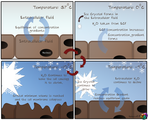
Osmotic Shock diagram

Surviving At Sub-zero Temperatures

To survive at freezing temperatures, organisms must combat the freezing process. Mammals and birds are endothermic, thermoregulators and thus produce their heat to stay warm. Thermoconformers, i.e. ectothermic organisms such as invertebrates and lower vertebrates, do not produce their own heat and must avoid freezing by other means. The body temperature of thermoconformers (Tb) is equal to the environment temperature (Ta) i.e. Tb=Ta. Problems generally occur when Ta < 0oC.

For example, freshwater fish have a body fluid freezing point of around -0.6oC. The freezing point of freshwater is around 0oC. Because the environment freezing point is higher than that of the body fluids it is unlikely the fish will be harmed by freezing.

However marine fish (still with a freezing point of around -0.6oC) face a bigger threat. This is due to the freezing point of seawater being lower, at around -1.38oC (due to a higher osmolarity as discussed earlier). This means the marine fish can freeze before their environment; therefore they must adapt different survival strategies.

To survive freezing conditions there are two options; freeze tolerance and freeze avoidance. Tolerance requires that the organism’s body learns to cope with the freezing process, whereas avoidance employs certain strategies to avoid freezing altogether.

These strategies include, super cooling and freeze point depression. Supercooling is widespread amongst arthropods; it involves the voidance of all ice nucleating agents from the body, thus allowing body tissue to supercool. An example of how this is achieved in insects is the emptying of the gastrointestinal tract. This prevents ice nucleating agents from entering the body. By entering hibernation at the same time it is possible to prevent the disturbance of bodily fluid and thus reduce the risk of freezing.

Freeze Point Depression as a Means of Survival

Utilising freeze point depression as a means of survival requires the regulation of body fluid freezing points. One method of achieving this is to increase the osmolarity of the body fluids which will decrease their freezing point. This technique is known as colligative defence. There are a few candidate molecules which could be used for colligative defence:

  • Electrolytes, such as salts and ions, however these can be damaging to the body at high concentrations as they can disrupt membrane potentials and proteins.
  • The other candidates are low mass organic solutes such as short chain sugars and alcohols; these have the benefit of not being harmful to the body at the concentrations required for freeze protection.

Colligative Defence

The typical molecule used for colligative defence is glycerol; such use has been demonstrated in Alaskozetes antarcticus, a species of mite which is able to survive in freezing temperatures. During the winter A. antarcticus cellular concentrations of glycerol are around 7 times higher than in summer. This rise in concentration of glycerol causes ΔFp (the change in freeze point depression) to fall drastically.

A 5M concentration of glycerol means a ΔFp of -9.3oC allowing survival at much lower temperatures. A fall in freezing point also means a fall in super cooling point too; a 5M concentration of glycerol will lead to a super cooling point of around -55oC.

Non-Colligative Freeze Point Depression

An alternative to the typical colligative defence is the use of non-colligative proteins which work in a different manner to prevent the formation of ice crystals. The proteins responsible for this are known as antifreeze proteins (AFPs) and antifreeze glycoproteins (AFGPs).

AF(G)Ps work by lowering the temperature at which ice crystals can grow, thus reducing the freeze point of body fluids. However they do not alter the melting point, a concept known as thermal hysteresis. This means that the inclusion of AFPs in the blood will lower the freezing point to around -2.0oC, but the melting point of the blood will not change, staying at around -0.9oC.

Under normal circumstances, the freeze point (Fp) is equal to the melting point (Mp). Colligative defence mechanisms decrease both Fp and Mp, but non-colligative defences, via AFPs, reduce the Fp leaving the Mp constant.

AFPs also differ from colligative defences in another manner. With colligative defence, the freeze point changes in a linear relationship to the osmolarity of the colligative molecule. However AFPs depress the freeze point greater than expected. They reduce the freeze point to the maximum possible at concentrations which would barely see a reduction in freeze point by typical colligative molecules.

AFPs inhibit recrystalisation, that is, they prevent smaller ice crystals joining together to form large ice crystal lattices. They do this by attaching to the, typically flat, growing planes of the ice crystal. By doing this, they cause highly curved fronts to form in the ice crystal. This disrupts the structure and prevents further water molecules adding to the lattice as doing so would require much larger amounts of energy than normal. This is known as the adsorption-inhibition model.

The production of AFPs may exhibit seasonality, for example, polar fish of the Antarctic (where there is a continuous ice sheet all year round) continuously produce AFPs. However polar fish of the Arctic (where seasonality is observed) only produce AFPs in the winter when they are at most risk of internal freezing. By doing this, they are able to conserve energy for the winter period (as the production of AFPs is energetically expensive).

The seasonal production of AFPs is controlled by the photoperiod (i.e. the day length). A long daylength stimulates the hypothalamus to produce GHRH (growth hormone regulation hormone) which in turn stimulates the pituitary gland to produce growth hormone (GH). Receptors in the liver for GH are activated by GH and induce a signal within liver cells. This signal inhibits the transcription of the AFP gene and thus no AFP mRNA is transcribed and no AFPs produced.

During shorter photoperiods (i.e. winter) instead of GHRH, the hypothalamus produces somatostatin (also known as growth hormone inhibiting hormone GHIH). This prevents the secretion of GH by the pituitary gland and thus liver cell GH receptors produce no signal. The AFP gene is not inhibited, produces mRNA and mature AFPs are produced and exported in to the blood.

Freeze Tolerance

Some organisms choose to cope with freezing by a different means, instead of trying to prevent freezing they actually induce freezing at ‘high’ temperatures so they are able to cope. This is widespread amongst insects as well as some intertidal invertebrates (e.g. barnacles), amphibia (e.g. green tree frog) and reptilia (e.g. garter snake).

Their method of coping with freezing is to control the formation of ice crystal (and thus physical damage). This allows them to freeze slowly and avoid super cooling (which would cause rapid freezing). They initiate freezing at higher than usual temperatures by not allowing body fluid temperatures to fall below freezing; they do this by ‘innoculative freezing’, a process where ice nucleating agents (INAs) are actively taken up from the environment. This prevents super cooling from occurring. However, freezing must only occur extracellularly, even freeze tolerant organisms cannot survive intracellular freezing.

An example of innoculative freezing is observed in the wood frog, this species of frog actively internalises ice crystals from its environment to act as INAs. It does this by pressing its body against ice which allows the thin membrane of the under belly to be penetrated by ice crystals. Another example is the crane fly, which instead of taking INAs from the environment, has its own protein ice nucleators which allow initiation of freezing at higher temperatures.

As discussed earlier, with osmotic shock, the formation of extracellular ice crystals causes the extracellular osmolarity to increase. The intracellular concentration equilibrates and, as a result of increased osmolarity, freeze point depression occurs. Normally this cycle of increased extracellular osmolarity and equilibration of the intracellular osmolarity would cause the cell to shrink to its critical minimal volume and rupture; however freeze tolerant organisms have developed ways to deal with these major changes in cell volume and osmolarity.

Cryoprotectants

Cryoprotectants are molecules found in freeze tolerant organisms which protect them against the cold. Cryoprotectants can be colligative or non-colligative:

  • Colligative – Colligative cryoprotectants require higher concentrations than non-colligative to function well. They work by accumulating in the intra- and extracellular fluids and limiting the amount of water transfer between them.
  • Non-colligative – Required in lower concentrations, non-colligative cryoprotectants work by binding to the cell membrane. Typically, as the cell osmolarity increases due to freezing, the cell membrane becomes more rigid as the phospholipids of the move closer together. Non-colligative cryoprotectants bind to the phospholipids of the cell membrane and prevent them from moving close enough to form a rigid structure, thus keeping the cell membrane as fluid as possible and preventing cell collapse.

The most common cryoprotectants are sugars and polyols (alcohols), they have the benefits of being non-toxic, highly soluble and metabolically inert. Often, multiple cryoprotectants are used, as opposed to relying on just one form. Examples of typical cryoprotectants are; glycerol, trehalose and sorbitol.

Cryoprotectants are synthesised from glycogen which means that to survive long, cold winters, the organisms must have large glycogen stores in the liver and fatty tissues. Two other considerations for survival are how to deal with the anoxic conditions endured during freezing and coping with the reactive oxygen species which occur during the thawing process.

Freeze Tolerance of the Wood Frog (Rana sylvatica)

The way in which the wood frog copes with freezing is to control ice formation in a manner that is slow and occurs from the periphery to the core. Freezing of the peripheral extremities causes signals to be sent to the liver to begin producing cryoprotectants. The glycogen stores begin to be broken down into glucose which acts as a cryoprotectant for the wood frog.

An advantage the wood frog has for surviving freezing are the large fluid filled spaces within the abdomen. The process of freezing in the wood frog is as follows:

  • Periphery
  • Large fluid filled spaces
  • Organs
  • Heart and liver

This is where the large fluid filled spaces are advantageous – when freezing begins to occur in the organs, ice is actively transported from the organs to the large fluid filled spaces limiting the amount of freezing which occurs in the organs. When the organs begin to freeze, there is minimal water content as the majority now resides in the large fluid filled spaces. These spaces are less likely to suffer from ice damage when compared to important organs, which is why the organs shift the water there.

The cryoprotectant, glucose, is transported around the body by the blood, but as the periphery begins to freeze, no further glucose can diffuse. However, as glucose is still being produced, the organs still receive glucose i.e. their concentration of glucose rises. As the body begins to freeze totally, the last places to receive glucose are the liver and heart, which means they have the highest concentration of glucose – this is important for the thawing process.

When completely frozen, breathing ceases as does the heartbeat however the frog continues its metabolism at a highly reduced rate. This keeps the frog alive and allows it to deal with the anoxic conditions.

When the wood frog eventually begins to thaw, it melts from the core to the periphery (the opposite direction to which it froze). This is because the highest concentration of glucose is found in the heart, liver and other organs. As glucose is a colligative defence molecule, at higher concentrations it reduces the freezing point, but it also reduces the melting point. Therefore, the organs with the highest concentration of glucose (liver and heart) have the lowest melting point and thus thaw first.

As the frog thaws from inside to out, it must cope with the readjusting volumes of cells and fluid filled spaces due to the ice melting. It must also readjust its metabolism from the reduced anoxic levels to increased normoxic levels. This involves the resumption of breathing and the heartbeat. One final concern is the presence of reactive oxygen species which occur during the thawing process, this requires the use of antioxidant molecules to inhibit oxidation of other molecules by the reactive oxygen species.

Bacterial Pathogens of the Respiratory Tract

Mannheimia haemolytica

M. haemolytica is responsible for causing contagious bovine pleuropneumonia, a bacterial disease which causes pneumonia and inflammation of the lung membranes. It is a Gram-negative coccobacillus (elongated, rod spheres) which shows mild haemolysis when plated on blood agar plates. This species comprises of 12 capsular serotypes (of which some are more responsible for disease than others). Serotype A2 is associated with sheep pneumonia.

Typically, diseases associated with M. haemolytica present themselves as fever, along with nasal discharge, coughing, inappetance and weight loss. Death associated with this bacteria is typically due to acute fibrous pleuropneumonia. Pathological observations would reveal an obstruction of the bronchioles with a fibrous exudate and an accumulation of neutrophils and fibrin in the alveoli. There will also be thrombosis and distension of the lymphatic vessels.

Mannheimia haemolytica as an Opportunistic Pathogen

M. haemolytica resides in the nasopharynx and tonsils of healthy cattle. A balance between the host and bacteria, under good environmental conditions maintains homeostasis between the host and bacteria (i.e. no disease). Should environmental conditions alter however, the balance may be tipped in favour of the bacteria, removing the status of homeostasis and resulting in disease. Key inciting events for disease include; weaning, adverse weather conditions, dehorning, feed changes and transportation i.e. causing stress. Stress provokes the bacteria in the nasal/tonsillar mucosa allowing them to be inhaled in to the lungs.

In healthy cattle clearance of the bacteria from the lungs is efficient enough to prevent disease. This suggests that stress induced alterations in immune functions can lead to host susceptibility and thus development of disease.

Close contact with other animals increases exposure to microorganisms which upsets the natural balance of commensal bacteria within the host. Changes in the natural microflora can cause M. haemolytica to revert to a pathogenic state. Slight changes in the environment are responsible for creating a favourable situation for M. haemolytica to colonies the lung and thus cause disease.

Mannheimia haemolytica Virulence Factors

Virulence factors of M. haemolytica include:

  • Adhesins – Required for initial colonization and adherence to host cells
  • Capsular polysaccharide – Helps prevent opsonisation and thus has anti-phagocytic properties
  • Sialoglycoprotease – An enzyme which cleaves IgG and thus reduces opsonisation of the bacteria
  • Neuraminidase – An enzyme which reduces the viscosity of respiratory mucus, reducing the chance of the bacteria being excreted via mucus
  • Iron-binding proteins – Removes iron from the host, for the benefit of the bacterium
  • Leukotoxin – A primary toxin involved in the pathogenesis of M. haemolytica

Mannheimia haemolytica Leukotoxin

Leukotoxin (LKT) is an actively secreted exotoxin which targets leukocytes, primarily neutrophils. The toxin is encoded for by four genes (lkt A-D) which can be found in the RTX toxin operon.

  • lktA encodes for the active toxin
  • lktB and lktD encode for the secretion of the toxin
  • lktC encodes for proteins responsible in the transportation and activation of the toxin

LKT help bacteria survive by allowing them to evade phagocytes. It is a type 2 exotoxin (membrane-damaging) which binds to leukocytes via the cell surface receptor CD18 (an integrin). It forms pores in the cell membrane of leukocytes which leads to an influx of K+/Ca2+ ions. This promotes swelling of the leukocyte and ultimately lysis of the cell.

Neutrophils affected by the toxin also become overly active, they overproduce certain mediators, reactive oxygen species and proteases, all of which promote further cellular and tissue damage within the host. Lesions develop which are filled with fibrous exudate and thrombosis of lymphatic vessels occurs. The alveolar epithelium also becomes damaged which is believed to be associated with neutrophil infiltration.

Controlling Mannheimia haemolytica Infections

Methods which could be implemented to reduce cases of M. haemolytica infections include:

  • Livestock management – Reduce stress of cattle by effectively managing when and how calves are weaned, sold and transported
  • Antibiotic use – Injectable antibiotic regimes are extensively used, especially in the intensive farming seen in large feedlots. However evidence shows that M. haemolytica are developing resistance to many of the common antibiotics used, such as; penicillin, ampicillin, tetracycline and sulphonamide
  • Vaccines – Both killed and live attenuated vaccines are used as well as cell-free supernatants such as the leukotoxin or capsular polysaccharide
  • Genetically Modified plants – A suggestion has been made for the use of GM crops to feed cattle in the future. The GM plants would produce M. haemolytica antigens which would essentially act as edible vaccines

These control methods could help to restore balance to the situation (previously unbalanced by negative environmental factors). Thus homeostasis between host and bacteria would be restored and disease prevented.

Bordetella bronchiseptica

Bordetella bronchiseptica is an evolutionary progenitor of B. pertussis and is one of the organisms responsible for causing kennel cough in dogs. Kennel cough is the term used to describe a disease which causes coughing in a dog due to inflammation of the trachea and lower airways. Although kennel cough is primarily caused by infection of the airways with B. bronchiseptica it can also be caused by viruses such as canine parainfluenza. Clinical signs of kennel cough include; an intense cough, mucus, nasal discharge and breathing difficulties.

Kennel cough gets its name from the frequency of infections which arise in dogs temporarily kenneled for example whilst the owner is on holiday. A number of dogs can carry the infection and the close contact in the kennels promotes transfer of the infection. Infection is caused either airborne transmission or via direct contact and has an incubation period of 3-10 days. Infected dogs can carry and shed the infection for up to four months after recovering from the disease. Because it require a only a few B. bronchiseptica bacteria to establish and infection in the airways (i.e. low infectious dose), B. bronchiseptica are considered highly infectious.

Bordetella Bronchiseptica Virulence Factors

Adhesins

  • Fimbriae – Associated with the initial adherence of the bacteria to epithelia. This form of adhesin allows the bacteria to latch on to host cells and begin to proliferate and form infectious colonies.
  • Filamentous Haemagglutinin Adhesin (FHA) – A large, filamentous protein which serves as a dominant attachment factor for adherence to host colliery epithelia cells of the respiratory tract. It is associated with biofilm formation and possesses at least four binding domains which can bind to different cell receptors on the epithelial cell surface.
  • Pertactin – An ‘autotransporter protein’ capable of getting to the cell surface without the need of accessory proteins. Pertactin also acts as an adhesin.

Adenylate Cyclase (cyaA)

  • Structurally similar to M. haemolytica leukotoxin
  • Different function, cyaA enters host cells and catalyses large amounts of cyclic AMP
  • This disrupts cell signaling pathways, impairing cellular function and inducing apoptosis whilst allowing the bacterium to avoid phagocytosis
  • It also inhibits the expression of interleukin 12 (IL12) an inflammatory cytokine

Dermonecrotic Toxin

  • A type 3 exotoxin which acts intracellularly
  • Precise mechanism is unknown however the overall effects on the cell include; actin reorganisation, increased DNA synthesis and binulcleation
  • Its overall contribution to the pathogenesis of B. bronchiseptica is currently unknown

Not all these virulence factors are active at the same time however, only some are active depending on the temperature. At 37˚C all are active except the flagellum, at at 27˚C none are active except the flagellum. What this shows is that there is a global regulation of gene expression depending on the requirements of the bacteria’s survival.

For example, when B. bronchiseptica is required to colonise the respiratory tract (indicated by a 37˚C temperature i.e. body temperature) it is not going to need to use its flagella for movement so the genes encoding for flagella motion are not expressed. In contrast when B. bronchiseptica is outside of its target destination e.g. the nasal passageway (indicated by a lower temperature) the flagella genes are expressed again as movement will be required to reach the target destination (the airways). Genes encoding for virulence factors will not be expressed as there is no need when the bacterium is not at its target site of pathogenesis.

Treatment of Kennel Cough

Kennel cough is susceptible to many antibiotics (it is however resistant to erythromycin). There are bivalent canine vaccines available which vaccinate against B. bronchiseptica and canine parainfluenza virus. Many of B. bronchiseptica outer proteins are highly immunogenic which makes good vaccines (e.g. fimbriae, FHA, pertactin)

In Summary

M. haemolytica and B. bronchiseptica are two important veterinary pathogens as they both inhabit the same ‘niche’. This is interesting because they also share similar virulence factors. Both diseases result directly from the disruption of the respiratory epithelium and from immunopathology and both diseases are often polymicrobial, involving mutliple agents.

Diagnosis relies on isolation and cultivation of bacteria on agar plates after which the correct antibiotic treatment can be given. Vaccinations are available for both diseases.

These diseases show how the management of animals can influence disease epidemiology.

Antibiotic Resistance

Introduction

The generic purpose of an antibiotic is to prevent the growth and/or survival of invading organisms whilst causing minimal damage and toxicity to the host. The typical mechanism of antibiotic action involves targeting specific enzymes or substrates of the invading bacterial species. Antibiotics may be either bacteriocidal (i.e. kill bacteria, e.g. β-lactam antibiotics) or bacteriostatic (i.e. slow bacteria growth and reproduction e.g. tetracyclines). However, the majority of bacteria are actually killed by the host immune system, the administration of antibiotics is typically only to aid the immune system and thus speed up recovery.

Some antibiotics may be synergistic, that is, when used with other antibiotics the overall therapeutic effect is greater than the sum of their individual effects, this allows for reduced doses to be administered.

Antibiotics can have either target a narrow spectrum of bacteria (such as the penicillins and macrolides), or a broad spectrum of bacteria (such as the aminoglycosides, cephalosporins, quinolones and some synthetic penicillins).

The use of antibiotics in food animals is subject to EU legislation which governs maximum residue limits (MRLs), the maximum concentration of antibiotic allowed to be administered to the animal for it still to be fit for human consumption. Antibiotics must be prevented from entering the food chain in this manner, so MRLs have been categorised into 4 annexes:

  • Annex 1 – MRL has been fixed
  • Annex 2 – MRL not required
  • Annex 3 – Provisional MRL
  • Annex 4 – No MRL can be set

The use of annex 4 antibiotics (e.g. chloramphenicol) is prohibited in animals destined for human consumption, with use of annex 1 or 2 antibiotics being preferred.

Choice of Antibiotics

There are many factors to consider when selecting an appropriate antibiotic for use. The first question to ask should be, ‘Is the infection bacterial?’ Antibiotics should only be administered for bacterial infections as they are of no use against viral infections. It is also important to remember that antibiotics only treat the infection and do not act as anti-inflammatories or anti-pyretics.

The site of infection is another consideration, as whilst some antibiotics will be of great use for skin infections, they may not be as useful for respiratory tract infections. The species of bacteria must also be considered, this will firstly require the culture and identification of the invasive organism to determine the species. However, within a species there may be variations of antibiotic susceptibility so it is important to generate a resistance profile i.e. determine which antibiotics the bacteria is resistant to and thus avoid prescribing these antibiotics. It must then be decided whether to use a broad or wide spectrum of antibiotic and wether this antibiotic should be bacteriocidal or bacteriostatic.

Bacteriostatic antibiotics will require a duration of therapy which gives the host cellular and humoral immune responses enough time to eradicate the bacteria. Alternatively bacteriocidal antibiotics are used when the host immune system is considered to be ineffective against the invasive organism.

Other factors to consider include:

  • The distribution of the drug throughout the body
  • The cost for a course of the antibiotic
  • The toxicity posed to the host
  • Any underlying disease which may be affected by the use of the antibiotic
  • Whether the host is pregnant or juvenile
  • The practically of the drug i.e. the route of administration, dosage, frequency of dose and the duration of action etc.

The Cascade

One final consideration to be made when administering antibiotics for animals is ‘The Cascade,’ which is a regulation governing restrictions on the administering of veterinary medical products.

In animals, an antibiotic must be licensed for use with a particular species and disease. However if no such antibiotic exists, to avoid unacceptable suffering, the veterinary surgeon may prescribe another antibiotic in order of the cascade:

  1. A veterinary medicine licensed for use in another species or in the same species but for a different use. This is known as ‘off-label’ use.
  2. A medicine licensed for use in humans.
  3. An unlicensed medicine, created and prescribed as a ‘one-off’ by the veterinary surgeon.

Determination of Resistance

Two important factors to consider when attempting to determine whether a bacteria is resistant to a particular antibiotic are:

  • The Minimal Inhibitory Concentration (MIC) – The minimum concentration of a drug which prevents the growth of bacteria
  • The Minimum Bacteriocidal Concentration (MBC) – The concentration of a drug which causes a 99% reduction in a bacterial innoculum over a given period of time.

Also of importance are:

  • Pharmacodynamics – The effect of a drug on a pathogen i.e. the MIC and MBC
  • Pharmacokinetics – The effect of the body on a drug i.e. the absorption, distribution, metabolism and excretion of the drug

Breakpoints are predetermined concentrations of an antibiotic, which determine whether a bacteria is deemed susceptible or resistant to that antibiotic. If this concentration of antibiotic causes a reduction in bacterial numbers or prevents further growth then that strain of bacteria is defined as susceptible to the antibiotic in question. If the bacteria does not at least show signs of prevented growth, the bacteria is defined as resistant to the antibiotic.

Disc Diffusion

One method of determining antibody resistance is disc diffusion. A species of bacteria is isolated and cultured on growth medium. After an incubation period that allows for visible colonies to be observed on the growth medium, diffusion discs are placed around the growth plate. These discs are impregnated with different antibiotics. After a short incubation period, susceptible bacteria around the discs will have been killed, indicated by a clearing in the colonies. The diameter of the clearing can be used to determine how susceptible the species is to an antibiotic. If there is no clearing (or a very small clearing) this indicates resistance to the antibiotic which impregnated the disc.

Mechanisms of Antibiotic Resistance

Intrinsic Resistance

Intrinsic resistance is that which is not acquired, inherent resistance, such as a natural low permeability to antibiotics due to a bacterial envelope. This type of resistance is characteristic to almost all representatives of a species. An example of this type of resistance is observed in Pseudomonas species. Their resistance to antibiotics is owed to the low permeability of their bacterial envelope to certain antibiotics and the presence of a multi-drug efflux pump.

This efflux pump occurs naturally in bacteria and usually removes waste products from the cell, such as bile, fatty acids and organic solvents. However a mutation in some species also permits the efflux of antibiotics from the cell thus preventing them from exerting their action upon the bacteria. This trait is also acquirable however, such as the efflux pump gene responsible for tetracycline resistance.

Acquired resistance

Antibiotic resistance genes found on plasmids, can be transferred between individual bacteria, hence non-resistant bacteria can easily acquire resistance. Acquired resistance can also occur due to mutations. Beneficial mutations can then be transmitted around the bacterial population. The biggest resistance problems occur in Gram-negative organisms due to the large numbers of plasmids found within the population, another resistance problem occurs in Staphylococcus species i.e. MRSA and MRSP.

Targets of Antibiotics

Antibiotics can work in a number of ways:

  • Inhibit cell wall synthesis e.g. penicillins, cephalosporins, carbapenems, glycopeptides
  • Inhibit DNA synthesis e.g. fluoroquinolones
  • Inhibit RNA synthesis e.g. rifampicin
  • Inhibit folic acid synthesis e.g. sulfonamides, trimethoprim
  • Inhibit protein synthesis e.g. macrolides, chloramphenicol, tetracycline, aminoglycosides

However, bacteria have a number of mechanisms to resist the effects of antibiotics:

  • Decrease permeability to the antibiotic
  • Inactive the antibody by chemically altering it with enzymes
  • Efflux of the antibody via efflux pumps in the membrane
  • Alter the target of the antibiotic, so it no longer has an effect
  • Bypass steps in metabolism which the antibiotic targets

How do Antibiotics Work?

β-Lactams

β-lactams can be both natural and semi-synthetic. They work by inhibiting enzymes associated with the synthesis of peptidoglycans, so essentially inhibit cell-wall synthesis. Such antibiotics include; penicillin and its derivatives, cephalosporins, monobactams and carbapenems.

However over use of these antibiotics has selected for bacteria which have developed resistance, in the form of β-lactamase – an enzyme which hydrolyses β-lactams. The hydrolysation of β-lactam antibiotics will alter their conformation and thus they will no longer have an antimicrobial effect.

To combat this, β-lactamase inhibitors are now used alongside β-lactam antibiotics. Although β-lactamase inhibitors do not act as antibiotics alone, coupled with β-lactams, they can effectively target bacteria. β-lactamase inhibitors, inhibit the enzyme responsible for hydrolyzing β-lactam antibiotics, thus when coupled with β-lactams, the antibiotics are free to act upon their targets again – enzymes associated with cell wall synthesis. An example of this is Synulox, which contains the β-lactam amoxicillin and the β-lactamase inhibitor clavularic acid.

Bacterial β-lactamase enzymes work by hydrolyzing the ring-bond of the β -lactam which denatures their structure thus preventing them from exerting their antibiotic action.

β-lactamases can be either chromosomally derived (typically in Gram-negative bacteria) or  their genes can be found on plasmids and thus spread throughout a population. Extended spectrum β-lactamases are encoded for on plasmids (plasmids; TEM, SHU and CTX-M). These extended spectrum β-lactamases can catalyse a broad spectrum of β-lactam antibiotics.

Other mechanisms for penicillin (and derived antibiotics) resistance include; alterations in the penicillin binding protein found in bacterial cell membranes. Alterations in this protein reduces the bacterial affinity for penicillin. Similarly, the acquisition of a novel penicillin binding protein (mecA) also reduces affinity for penicillin. This gene is found in MRSA and is what makes it so resistant to penicillin its derivatives.

Aminoglycosides

Aminoglycosides irreversibly bind to the 30S ribosome and freeze the initiation complex. This effectively inhibits protein synthesis within the bacterium. Aminoglycosides have a broad spectrum but do not target anaerobes. They can also have a synergistic effect when used with β-lactams. They do however pose a toxicity risk; nephrotoxicity (toxic to the kidneys) and ototoxicity (toxic to the ear, specifically the cochlea or auditory nerve).

The mechanisms for resistance against aminoglycosides include; alteration of the bacterial ribosomes which prevent aminoglycosides from binding, decreased permeability to the antibiotics or the inactivation of aminoglycosides by mechanisms such as acetylation, phosphorylation and adenylation. These mechanisms are facilitated by aminoglycoside enzymes found in the bacteria.

Tetracyclines

Tetracyclines are bacteriostatic and similarly to aminoglycosides, they bind to the 30S ribosome (however, they bind irreversibly). They also inhibit RNA from binding to the 70S ribosome and thus inhibit protein synthesis. They have a broad spectrum of targets, but there are also many tetracycline resistant strains of bacteria.

Fluoroquinolones

Fluoroquinolones inhibit DNA synthesis, they do this by targeting the DNA enzymes gyrase and topoisomerase. They too have a broad spectrum, but bacterial plasma-mediated QNR genes provide resistance against the antibiotic. QNR genes offer low level protection for gyrase and topoisomerase from fluoroquinolones, however, the presence of QNR genes increases the mutation rate of gyrase and topoisomerase genes. Mutation of these genes can further increase resistance to fluoroquinolones.

Trimethoprim

Trimethoprim inhibits the bacterial dihydrofolate reductase (DHFR) enzyme which therefore inhibits folic acid synthesis. Again, trimethoprim has a broad spectrum, it is often used in combination with other antibiotics.

Resistance to trimethoprim occurs if the DHFR enzyme becomes less susceptible to the antibiotic e.g. through mutation, or if the metabolic step in folic acid production which requires DHFR is skipped altogether thus removing the target for trimethoprim completely.

Antibiotic Resistance

Resistance has always existed, even before antibiotic use. However the use of medical antibiotics have increased the prevalence of resistance. It is important to note though, that antibiotics do not cause resistance. They do however select for resistance already prevalent in a population.

Staphylococci Virulence

Introduction

Staphylococcus is a gram positive, cocci shaped, genus of bacteria. Observed under a microscope will reveal they exist in microscopic ‘grape-like’ clusters. One species of staphylococci, Staphylococcus aureus, can grow at temperature ranges of 15-45ºC and at  a relatively high NaCl concentration of 15%.

Catalase Test

One of the main tests used to differentiate between bacterial species is the catalase test. This test determines whether or not the enzyme catalase is present. Catalase, is responsible for the breakdown of hydrogen peroxide. The test consists of adding a bacterial colony to a drop of hydrogen peroxide, if the bacteria is catalase-positive (i.e. has the catalase enzyme) bubbles of oxygen will be produced. This will not occur in catalase-negative bacteria. Staphylococci species are catalase-positive.

Oxidase Test

The oxidase test, is another major test used to differentiate between bacteria. It tests to see if certain cytochrome c oxidase enzymes are present. These enzymes are involved with the electron transport chain. There are a number of ways to perform this procedure, but they all typically involve adding a reagent to the sample bacteria and observing for a colour change. Typically the development of a blue colour indicates a positive result (oxidase enzymes are present), with no colour change indicating a negative result. Staphylococci species are oxidase-negative.

Coagulase Test

The final major test used for differentiation is the coagulase test. This test determines whether the enzyme coagulase is present, an enzyme responsible for the formation of blood clots and primarily associated with staphylococci species. However, within the Staphylococcus genus, there are both coagulase-positive and coagulase-negative species. S. aureus is an example of a coagulase-positive species, and S. epidermidis is an   example of a coagulase-negative species. The procedure involves adding blood plasma to the test sample, the development of agglutination after a short period of time indicates a positive result, with no agglutination indicating a negative result.

Staphylococci & Animals

Currently, around 34 species of staphylococci have been identified, with many of these being found in multiple species of animals. Different species of Staphylococcus have different preferred hosts, Staphylococcus tend to co-evolve with their hosts, but they are still able to cross species barriers. The major Staphylococcus found in dogs and cats are:

  • S. intermedius
  • S. felis

S. aureus is not commonly found in dogs and cats, it is found more often in other domesticated species however.

Staphylococci can be found in certain locations all over the body where they result in no disease. These are known as resident populations and include; mucosal surfaces, mucocutaneous junctions (such as the lips, nostrils, vagina, etc.) and the ear canal.

Staphylococci of Veterinary Importance

Coagulase positive:

  • S. aureus
  • S. intermedius
  • S. hyicus

Coagulase negative:

  • S. sciuri
  • S. equorum
  • S. epidermidis

Staphylococcus pseudintermedius

S. pseudintermedius is most commonly found on the skin of domestic dogs and cats. This species is zoonotic and thus has the potential to infect humans, although human cases of infection with this species are rare. Of increasing concern is the rise in human S. pseudintermedius infectious, particularly because of the resistance to antibiotics shown by this species.

Some clinical signs associated with infection of this bacteria are; otitis externa (inflammation of the outer ear), mastitis, infective endocarditis (inflammation of the inner layer of the heart), abscess formation, infection of wounds and primarily, chronic pyoderma (an inflammatory skin disease).

Alterations of the skin’s micro-environment can promote the growth of bacteria such as S. pseudintermedius. For example, inflammation provides humidity and warmth, both of which promote bacterial multiplication. Any trauma caused to the skin will further reduce epidermal defenses. Continuing with the inflammation example, irritation may cause the the animal to scratch intensely which can lead to damage of the upper layers of the epidermis and make infection for S. pseudintermedius easier. Any allergic skin diseases, or underlying immunological disorders will also contribute to the degree of infection.

Pyoderma

Pyoderma is a bacterial skin disease, caused by bacteria normally found on healthy skin (commensal) such as Staphylococcus. Because these bacteria are opportunistic, if skin becomes diseased or damaged, they may proliferate which can cause problems. Whilst pyoderma is primarily caused by underlying skin problems, it is also possible for pyoderma to occur on healthy skin, however this often indicates underlying immune system problems.

Pyoderma becomes a problem when commensal bacteria breach the epidermis and begin to proliferate and adhere to keratinocytes – the predominant cell type of the epidermis. Areas prone to infection are those where the skin creates folds which reduces air circulation and provides a warm, humid environment, perfect for bacterial growth.

Treatment of pyoderma typically involves the use of antimicrobials either topically or systemically for around 8 weeks. Whilst this may remove the initial pyoderma, it is also important to treat the underlying conditions responsible for the outbreak as reinfection may occur. Reinfection will require more treatment, which is of concern as this can lead to antimicrobial resistance. Methicillin-resistant Staphylococcus pseudintermedius (MRSP, i.e. similar to MRSA) has recently been identified.

Staphylococcus hyicus

S. hyicus is responsible for causing exudative epidermitis, an oozing inflammation of the skin, also known as ‘Greasy Pig Disease’. It occurs when abraded skin is invaded by S. hyicus bacteria which then cause infection. Lesions begin to develop after infection, which then spread to the hair follicles (known as folliculitis). Inflammation soon follows this causing erosion and ulceration and the lesions continue to growth, engulfing large amounts of the skin surface. Alongside this, the sebaceous glands will produce a black, greasy, exudate.

Whilst this disease can be treated with antibiotics, death often occurs as a result of starvation of dehydration, so it is important to offer the infected pigs electrolytes by mouth to ensure a steady recovery.

Virulent strains of S. hyicus produce an ‘exfoliative toxin’. A toxin responsible for the epidermal necrolysis. Isolation of this toxin and reintroduction into a healthy pig will interestingly, reproduce the observed disease.

Exfoliative Toxin

The histopathology of S. hyicus induced exudative epidermitis, is very similar to that of S. aureus scalded skin syndrome. A disease which causes fluid filled blisters to appear on the skin of humans. Their similarities are owed to the exfoliative toxins, A & B which they secrete. These toxins cause a detachment of the epidermal layer and result in the observed lesions and deformations of the skin. This is beneficial to the bacteria as it allows them to further penetrate and proliferate beneath the skin.

Staphylococcus aureus Diseases

Staphylococcus aureus is responsible for a wide array of diseases, these include:

  • Superficial lesions
    • Skin and soft tissue infections
    • Mastitis
  • Invasive infections
    • Osteomyelitis – inflammation of the bone marrow
    • Endocarditis – inflammation of the inner layer of the heart
    • Pneumonia – inflammatory condition of the lungs
    • Septicaemia – The presence of pathogenic organisms in the blood which can lead to a body-wide inflammatory state
  • Toxinoses
    • Food poisoning
    • Scalded skin syndrome – Formation of fluid filled blisters on the skin
    • Toxic shock syndrome – A potentially fatal disease caused by a S. aureus toxin

Virulence & Virulence Factors

Staphylococcus species can express virulence in a number of ways, these include:

  • Adherence and surface associated proteins
  • Bacterial capsules which prevent phagocytosis
  • Exoenzymes, extracellular enzymes secreted by bacteria
  • Proteases, enzymes which break down proteins e.g. nuclease
  • Exotoxins, toxins secreted by the bacteria

Adhesion

Bacterial adhesion to host cells is the first step in colonization. Adhesion typically requires the presence of bacterial adhesins. These include pili, fimbrae, the flagella or the cell surface itself. These surface proteins allow for the attachment to host cells, in particular the host proteins laminin and fibronectin. Adhesins which promote attachment to collagen fibres can lead to osteomyelitis (infection of bone marrow) and arthritis.

Clumping Factor

Clumping factor is a protein which binds to fibrinogen, it is responsible for the formation of blood clots and it what causes the agglutination in the coagulase test. Clumping factor also helps to evade the immune system, by coating the itself in fibrinogen the bacterium avoid opsonisation and thus phagocytosis.

Protein A

A surface associated protein found in S. aureus, it binds to the Fc regions (tail regions of antibodies, which bind to Fc receptors on cell surfaces) of immunoglobulins, particularly IgG. This causes incorrect orientation of the bacteria in relation to the IgG, this is beneficial  because it disrupts opsonisation and thus phagocytosis. Protein A can also bind to IgM associated with B-cells (the lymphocytes associated with the humoral immune response), this induces apoptosis in these cells and thus causes depletion.

The gene which encodes for this protein is the spa gene, this gene is often used for molecular typing of Staphylococcus.

Capsular Polysaccharides

Capsules are bacterial structures which encase the entire bacterium, they are useful for protecting the bacterium against phagocytosis but they can also be a virulence factor. The presence of certain capsules can act as a potent abscess potentiator, isolation and injection of such a capsule can produce sterile intra-abdominal abscesses.

Super Antigens

Normal antigens are able to generate an immune response which induces a reaction from only the appropriate cells of the immune system. Typically much less than 1% of body T-cells (the lymphocytes associated with cell-mediated immunity) are activated. A super antigen on the other hand, generates a much more potent immune response. It is believed super antigens can activate up to 20% of the T-cells in the body.

Such a large immune response actually works in favour of Staphylococcus species. It is thought that these bacteria can produce more than 20 types of super antigens, in the form of enterotoxins and exotoxins. Enterotoxins specifically target the intestines and can cause diarrhoea and vomiting. In severe responses to super antigens (such as the exotoxins) the host can develop Toxic Shock Syndrome (TSS). TSS is where the body cannot cope with the large amounts of inflammatory cytokines released by the T-cells due to the binding of the super antigen, this can lead to shock and multiple organ failure.

The vast majority of TSS cases are caused by S. aureus and S. pyogenes. The most potent inducer of TSS however is Toxic shock syndrome toxin 1, a toxin produced by S. aureus which is responsible for around 75% of all TSS cases. Again, this leads to over stimulation of T-cells and a drastic systemic release of inflammatory cytokines which leads to lowered blood pressure, fever and in severe cases shock and organ failure.

Exfoliative Toxins

Another virulence factor of staphylococci is their ability to produce exfoliative toxins, exfoliative toxin A and B (ETA & ETB). These toxins result in blister formation at the epidermis surface which allows the bacteria to further penetrate and spread beneath the skin.

Membrane Damaging Toxins

There are three main forms of membrane damaging toxins (also known as type-II exotoxins) excreted by Staphylococcus, these are:

  • α-toxin – A potent membrane damaging toxin which is important for tissue invasion. In humans, platelets and monocytes are particularly sensitive to it. Cells susceptible to the α-toxin have specific receptors which it binds to, allowing it to cause damage.
  • β-toxin – This membrane damaging toxin targets membranes rich in lipids. A classic test to determine whether β-toxin is present, is to plate a sample on sheep erythrocyte-enriched growth medium. If β-toxin is present, it will lyse the erythrocytes which will be apparent on the growth medium as a clear area (as opposed to red).
  • Panton-Valentine Leukocidin (PVL) – This membrane damaging toxin, targets the cell membrane of leukocytes for which it has a high affinity. It can cause skin and soft tissue infections as well as pneumonia. Its structure allows for the formation of a pore through membranes, causing leukocyte cell contents to leak out.

MRSA – Methicillin resistant Staphylococcus aureus

Methicillin resistant S. aureus are bacteria which have developed resistance to beta-lactam antibiotics – those which target cell wall synthesis of bacteria. Such antibiotics includes the penicillin family of drugs. Their resistance is conferred by the gene meAc, which encodes for the protein penicillin-binding protein 2a (PBP2)

To determine whether a species of S. aureus is resistant:

  • The sample is first cultured
  • It must then be identified as S. aureus
  • The next step is to determine wether or not the strain is methicillin resistant
  • Once resistance is confirmed, the strain is then typed

Samples should be obtained from typical sites of carriage which include; the nasal passageway, skin, perineum and faeces. To culture MRSA, the growth medium must be selective i.e. contains a beta-lactam antibiotic, so only MRSA will grow.

At-risk patients include:

  • Dogs/Cats – Recent surgery, presence of non-healing wounds, recent or current treatment with multiple courses of broad spectrum antibiotics.
  • Horses – As above, also any length in-patient hospitalization

Evolution of Bacterial Virulence

Point Mutations

Much like humans, bacteria are subject to evolution. Evolution occurs through beneficial mutations in the DNA. Because bacteria have much shorter generation times than humans, the process of evolution occurs much more rapidly. An example of how bacteria evolve through mutation is point mutation.

Point mutation is the random mutation of DNA nucleotides which occurs due to incorrect replication of DNA. The types of mutation that can occur are substitution of a nucleotide, deletion of a nucleotide or the addition of a nucleotide. For example, in a sequence of DNA a fragment of which codes for something (such as a protein) consists of multiple codons (sections of 3 nucleotides) if one of these nucleotides is altered in a point mutation, the codon (which codes for a specific amino acid) will be altered. Some alterations of codons will mean they encode for a different amino acid:

Initial DNA:                  … … TTA … …

After point mutation: … … TAA … …

In this case the selected codon in the initial DNA encodes for the amino acid leucine, however after the mutation, it no longer encodes for an amino acid at all as TAA is a stop codon (this closes the reading frame, meaning the rest of the gene is not read).

Point mutations may sometimes have no effect however, as multiple codons can encode for the same amino acid, for example:

Initial DNA:                  … … GCT … …

After point mutation: … … GCA … …

Both GCT and GCA codons encode for the amino acid alanine, so there are no alterations to the final protein which is encoded for by this gene.

Horizontal Gene Transfer

Bacteria are also able to ‘evolve’ by means other than mutation. Horizontal gene transfer is the sharing of genetic information between individuals in the population. Typical gene transfer occurs vertically i.e. parent to offspring, however with horizontal gene transfer genetic information can be spread to completely unrelated individuals.

There are three primary forms of horizontal gene transfer, these are:

  • Transformation – DNA is taken up from the environment
  • Conjugation – DNA is transferred directly between individual bacteria
  • Transduction – DNA transfer is mediated by bacteriophages (a virus which infects bacteria)

Transformation

Many bacteria are naturally transformable and have various means of DNA uptake from their environment. Bacteria able to do this have genes which encode for protein machinery which transports the DNA across the cell membrane from the environment. It is then possible for this DNA to be incorporated into the bacterial chromosome.

This ability has important implications in bacterial pathogenicity for example; Neisseria gonorrhea are able to uptake DNA from the environment which enables them to undergo possible conformational changes that make them more pathogenic. Another example would be Streptococcus pneumoniae and its possibility to acquire penicillin resistance.

Conjugation

Conjugation is the direct transfer of DNA between bacteria, this is of major advantage as advantageous genes (such as those which encode for antibiotic resistance) can be easily spread throughout the population.

DNA is transferred in the form of a plasmid (a circular DNA structure) in a unidirectional manner, that is only the donor requires the plasmid transfer-associated genes (i.e. those which encode for plasmid transfer mediating pili) for horizontal transfer via conjugation to occur.

Plasmids

Plasmids are typically circular in structure and consist of double stranded DNA. They replicate independently of the bacterial chromosome and are located in the cytoplasm, they are usually much smaller than the bacterial chromosome ranging from 1-100kb long. The genes found within the plasmid can be expressed by the bacterium and so proteins encoded for by the plasmid can be formed in the bacterium.

The most common plasmids are those which encode for antibiotic resistance. They are highly prevalent because of the great advantage they give to bacterial survivability. The are multiple genes found on plasmids which promote antibiotic resistance all with different mechanisms of producing resistance.

For example β-lactam antibiotics (such as penicillin derivatives) work by inhibiting cell wall synthesis, however β-lactamase is a bacterial enzyme which breaks the ring structure of these antibiotics are removes their antibacterial properties. These enzyme is encoded for by genes found within plasmids, which can easily be spread around the population thus increasing the resistance of the population to β-lactam antibiotics.

Other forms of plasmid-conferred attributes include:

  • Bacteriocins – Proteinaceous toxins which inhibit the growth of competitor species a specific example of this is colicin, a bacteriocin produced by Escherichia coli which exerts a cytotoxic effect within competitor species’ cytoplasm.
  • Toxin production – Toxins which affect other species such as the heat-labile enterotoxin produced by E. coli which causes diarrhoea or the Clostridium tetani tetanus toxin, the neurotoxin responsible for causing tetanus.
  • Protein delivery systems – Such as the Yersinia pestis secretion system, which allows them to inject protein materials into other organisms (such as immune cells), these injected proteins are known as YOPs (Yersinia outer proteins).
  • Adhesins – Typically pili (protein surface projections) which allow adherence to bind to cells, which is important in pathogenicity. Such adhesins can be found in E. coli and Shigella.
  • Metabolic activities – For example, the ability of Salmonella species to ferment lactose

Transduction

Transduction is the transfer of DNA which is mediated by bacteriophages (viruses which infect bacteria, combining their DNA with the bacterium host). Many bacteriophages have both lytic and lysogenic cycles of reproduction (typically viruses are one or the other). The lytic cycle is where the virus enters the cell, replicates using the cell’s own machinery and then lyses the cell releases large numbers of virus. The lysogenic cycle is where the virus incorporates its genetic material into the cell where it lies dormant until a later date, the viral genes are replicated when the cell replicates allowing them to reactivate and continue into a lytic cycle (formation of phages).

Bacteriophage-encoded virulence factors include:

  • Streptococcus pyogenes toxin (which causes scarlet fever)
  • Corynebacterium diphtheriae toxin
  • Clostridium botulinum toxin

E. coli O157:H7 contains around 1.4Mb of extra DNA when compared to E. coli K12 (a much less pathogenic strain which is used in laboratories). It is suggested that around 50% of this extra genetic material has been derived from bacteriophages.

Vibrio cholera Toxin

Another bacteriophage encoded virulence factor is the Vibrio cholera toxin, however for this toxin to be produced by the bacterium, genetic material from two bacteriophages must be acquired.

The initial bacteriophage (VPI phage) infects a non-toxic Vibrio cholera bacterium, its transduced genetic material encodes for pili proteins which act as an attachment site for the secondary bacteriophage (CTX phage). When CTX phage binds, the genetic material it incorporates encodes for the production of the cholera toxin and thus the bacterium becomes toxogenic.

Recombination

Transformed and transduced (i.e. non-plasmid DNA) is inserted into the recipient bacterial chromosome by recombination. Recombination is also responsible for the rearrangement of DNA fragments around the genome (genomic plasticity) which enables the bacteria to survive in rapidly changing environments due to the alterations in the genes which are expressed. Recombination can be either homologous (RecA dependent – a DNA repair and maintenance protein) or non-homologous.

Homologous Recombination

DNA inserted into the bacterium can be incorporated into the bacterial chromosome if there are regions of homology expressed on both the foreign transmitted DNA and the bacterial chromosome DNA. The differences between the foreign and bacterial DNA will cause RecA to attempt to ‘repair’ the chromosome. In doing so, the foreign DNA may be incorporated into the bacterial chromosome, (there is also the chance that it will be disregarded).

Non-homologous Recombination

Non-homologous recombination allows genetic material to enter the bacterial chromosome without RecA. Small fragments of DNA known as insertion sequences (ISs) are what enter the chromosome. ISs typically consist of DNA fragments from bacteriophages or elsewhere, however, all that an IS codes for is its motility and insertion. ISs integrate into the bacterial chromosome through self-encoded transposition enzymes (as opposed to RecA) and when within the chromosome they facilitate replication of themselves. The short ISs then become numerous within the bacterial chromosome.

The presence of multiple ISs within the bacterial chromosome is what allows genetic plasticity (The movement or deletion of short fragments of DNA within the chromosome). Genetic plasticity leads to alterations in genes or the removal of genes altogether, this has the potential to increase pathogenicity. For example, Bordetella bronchiseptica is a gram negative bacterium responsible for causing infectious bronchitis, which rarely infects humans. B. pertussis on the hand is the major causative agent of pertussis (whooping cough) and is an obligate human pathogen. It is believed that genetic plasticity caused a reshuffling of the B. bronchiseptica chromosome which lead to the formation of the obligate human pathogen B. pertussis.

Transposons

Transposons are transmitted by similar means to ISs, however they are more structurally complex and much larger, due to the greater amount of genetic information they carry. This extra genetic material encodes for attributes such as antibiotic resistance, toxins and enzymes.

Some transposons (known as conjugative transposons) excise themselves from the bacterial chromosome to form circular intermediates of DNA, much like plasmids. These can also be transmitted around a population, again like plasmids, to spread resistance to antibiotics etc.

Pathogenicity Islands

Pathogenetic islands are also laterally-transmitted. They are large integrated DNA elements which carry multiple virulence-associated genes such as those which encode for adherence factors, secretion systems, toxins and invasion factors. A bacterium may have multiple pathogenicity islands and due to the incorporation of multiple virulence factors on one island, the transmission of one island often causes a non-toxogenic bacterium to become pathogenic.

They may be incorporated into the main bacterial chromosome or any plasmids which may be present in the cytoplasm, the DNA found within these islands may contain remnants from bacteriophages and IS elements. The GC-content (guanine-cytosine content) is often different from the rest of the genome.

Diagnostic Tests Used for Viruses

Haemagglutination Assay

Introduction

A simple and rapid method of quantifying the amount of virus in a sample. Haemagglutination is the agglutination of red blood cells. Viruses with envelops or surface proteins are able to bind to the sialic acid, N-Acetylneuraminic acid found in the cell membrane of red blood cells. Because each agglutinating molecule (in this case each viral molecule) can bind to multiple red blood cells, a clump of cells begins to form, this is agglutination. This agglutination forms a lattice structure.

Haemagglutination Inhibition

Haemagglutination inhibition is the addition of an inhibitor of the virus. Antisera (a blood serum which contains antibodies) is used in this case. The antibodies will bind to the virus and thus prevent haemagglutination.

By creating multiple, increasing levels of dilution e.g. 1:2, 1:4, 1:8, etc. of virus to antiserum we can determine a haemagglutination titre, this is the highest value of dilution (i.e. smallest amount of virus) which inhibits agglutination.

A typical haemagglutination inhibition assay looks like this:

You can see that point of agglutination, where the contents of the well go from a homogenous cloudy red, to containing a red spot which is the agglutinated red blood cells. In the above example, sample E (the highlighted row) has a haemagglutination titre of 1:512 as this is the largest dilution which prevents haemagglutination.

By monitoring the haemagglutination titre, we can determine how the virus is progressing over time, for example:

  • 1st assay – an endpoint is reached at a dilution of 1:4
  • 2nd assay – an endpoint is reached at a dilution of 1:16
  • 3rd assay – an endpoint is reached at a dilution of 1:8

From this would we be able to see initially the virus count is low as it is inhibited at a low dilution, however at the next assay the haemagglutination titre is higher (greater dilution of virus reached before it is inhibited) this must mean the virus is replicating as the initial count of virus is higher (i.e. takes greater amount of dilution to reach inhibition). WIth the final assay, we can see that because the haemagglutination titre has decreased, the virus must be reducing in numbers, indicating recovery.

ELISA

Introduction

ELISA is an acronym for Enzyme-linked immunosorbent assay and is used to detect whether or not a certain antibody or antigen is present in a sample. To indicate the presence of an antibody or antigen, reporter molecules are used which are identifiable by a change in colour. Therefore, if an antigen or antibody is present, a colour change will be observed.

Method

A typical ELISA requires the use of a 96-well microtitre plate (as used in the haemagglutination assay), but the wells of the plate are coated with a known or unknown antigen. When diagnosing viruses, the antigen will be unknown as it is the sample which we are testing. To this an antiserum is added, if the virus antigen is present in the sample, then the antibodies will bind. If not, no binding will occur.

To determine whether or not an antibody-antigen complex has formed (i.e. a positive diagnosis of the virus), an antiglobulin is added. This is an antibody which binds to the antibodies used in the initial serum. For example, Goat anti-rabbit IgG is a rabbit antibody (Immunoglobulin G [IgG]) which will bind to goat antibodies. There are many possible variations of antiglobulins.

The antiglobulin will be ‘labelled’ with an enzyme (an enzyme is attached to the antibody which has a negligible effect on its binding capabilities).

Finally, the substrate of the enzyme using to label the antibody is added. This substrate will be broken down if the secondary antibody (antiglobulin) bound to the primary antibody. The break down of the substrate will be coupled with a colour change, for easy identification that this has occurred. The greater the intensity of the colour change, the higher the concentration of the initial antigen.

Sometimes a spectrophotometer (a device used to detect light intensity) will be used to determine the degree of change in colour, as this can be used to calculate reasonably accurate values for initial antigen concentration and thus virus concentration in the host.

Below is an example of the variations in colour which occur during an ELISA test. The coloured wells indicate positive samples:

This type of ELISA is known as an indirect ELISA test, however there are other forms of this test, such as sandwich ELISA, which is where the well is coated in antibody, to which an antigen binds and to this another antibody is added. The antiglobulin tests for the secondary antibody.

There are also competition ELISA tests, where the added sample is an antibody-antigen complex, this is added to antigen coated wells. If the concentration of initial antigen in the sample is high then there will be fewer available antibodies to bind with the antigen in the wells. The wells are washed to remove unbound antibodies and as with the indirect assay, enzyme-coupled antiglobulins are added with substrate that elicit a colour change. However in this case a high initial concentration of antigen in the sample will yield a low change in colour.

Leptospirosis

Introduction

Leptospirosis is a zoonotic disease caused by the bacterial genus Leptospira. Leptospires are spirochetes, a group of Gram-negative bacteria with long, thin, spiral structures and an internal flagella used for movement. The size of a typical leptospire is around 0.1μm wide and 6-20μm long. This narrow, helical structure enables them to burrow in to tissues, within tissues they may adopt a more spherical or granular appearance.

Etiology

The primary pathogenic strain of Leptospira is Leptospira interrogans, however there are also non-pathogenic strains such as Leptospira biflexa which is an environmental saprophyte (i.e. consumes dead organic matter).There are currently around >16 species of identified Leptospira.

Serovars & Serogroups

Serovars are groups of organisms, categorised depending on the antigens they present on their surface. Therefore if a number of organisms within a Leptospira species share the same antigens on their surface, they will be grouped together into one serovar. Differences in surface antigens occur within the same species for example, within the pathogenic L. interrogans species, around >250 serovars have been identified worldwide.

Serogroups are clusters of serovars, as there may be hundred of serovars within a species, it is useful to group those together which share similar properties. Important serovars of L. interrogans include:

  • Canicola (Primary reservoir host – Dog)
  • Icterohaemorrhagiae – (Rat, mouse)
  • Bratislava – (Rat, pig, horse)
  • Pomona – (Cattle, pig, skunk)
  • Grippotyphosa – (Rodents)
  • Hardjo – (Cattle)

Each serovar is not limited to its primary reservoir host however. They be transmitted to incidental hosts fairly easily. Incidental hosts include humans, dogs and cats as well as other domesticated animals.

Different serovars are also responsible for different clinical conditions which can range from abortion to haemorrhagic disease. An individual serovar may also cause different clinical conditions in different species, for example L.interrogans serovar hardjo causes abortion and still births in cattle, but in humans it can cause an influenza-like illness or liver/kidney diseases.

Epidemiology

Transmission Cycle

The transmission cycle of a typically Leptospira species is as follows:

  • Rodents shed Leptospira in their urine
  • Direct transmission of Leptospira to humans may occur at this stage
  • The urine contaminates the environment (e.g. soil, water) with Leptospira
  • Indirect transmission to humans may occur at this stage
  • Leptospira may be transmitted to other domestic animals via the environment
  • These animals may become infected and can shed Leptospira in their urine which can lead to direct transmission to humans or contaminate the environment as before

Rodents can acquire Leptospira from the urine contaminated environment, thus creating a cycle of transmission

Transmission via direct contact usually occurs by urine which contains the Leptospira organisms. However direct transmission may also occur via veneral or placental transfer as well as bite wounds or the ingestion of infected tissue material. Crowding of animals (such as in kennels or intensive farming) will enhance transmission of Leptospira. Animals which recover from the disease, may still be infected, thus making them carriers of Leptospira which can still be excreted chronically in their urine. This continues the spread of infection

Transmission via indirect contact can also occur. Methods of indirect transmission generally requires exposure to contaminated sources such as; soil, food, bedding or water sources. The bacteria enters susceptible hosts from the contaminated source via damaged skin or exposed mucous membranes such as in the nose, mouth, eyes etc. Leptospira remains viable in the environment (still able to cause infection) for months, this further enhances transmission.

Environmental Factors

The optimal habitat for Leptospira depends on their environment, if aquatic, optimal conditions are stagnant or slow moving waters. If terrestrial, a neutral or slightly alkaline soil pH is preferred. However, organisms may survive transiently in undiluted acidic urine. A typical temperature range of 0-25C is preferred, this often leads to seasonal fluctuations in the incidence of Leptospirosis.

Pathogenesis

Leptospira enters the host by penetrating mucous membranes via vunerable areas such as damaged skin, eyes, nose or the mouth. Their helical shape and flagella aids in tissue penetration. Upon entering the blood system, they begin to multiply rapidly. The presence of bacteria in the blood is called bacteraemia. They are then distributed around the body via the blood stream.

Once distributed around the body, they then further replicate in target organs and tissues (including the kidney, liver, spleen, central nervous system, eyes and genital tract). The incubation period is around 7 days, this factor depends on the species and the strength of the host immune system however.

The initial immune response will usually remove all Leptospira organisms from the blood and tissues but some will persist in the kidney tubules where they can continue to replicate. The Leptospira organisms in the kidney tubules manage to evade the host immune response by avoiding phagocytosis.

The damage done to the host’s organs and tissues is variable and depends on the virulence of the Leptospira serovar and how susceptible the host immune system is. The most serious of diseases occur in the incidental hosts, i.e. not the primary reservoir host.

Overview of Pathogenesis

Diagnosis

At present there are three different methods of leptospirosis diagnosis:

  • Detect leptospire antigens – Leptospire antigens will induce agglutination of antibodies. This can be tested using a microscopic agglutination test (MAT).
  • Isolation of Leptospires – Leptospires are isolated from the urine or infected tissues. However this can be very labour and time intensive as Leptospira species are slow to culture using growth medium, meaning it can take weeks before a positive/negative result is returned. Despite this, this method of diagnosis is probably the most reliable.
  • Polymerase Chain Reaction (PCR) – Molecular methods of diagnosis (such as PCR) are gaining popularity for diagnosing Leptospirosis, however PCR is unable to distinguish between serovars.

Prevention

For dogs there are currently two forms of vaccine available:

  • In the UK, a bivalent vaccine is used which protects against two serovars – canicola and icterohaemorrhagiae.
  • In the USA however, a quadrivalent vaccine is used, this protects against four serovars – grippotyphosa and pomona as well the canicola and icterohaemorrhagiae which the bivalent vaccine covers.

Some preventative measures are also being taken in cattle to protect against Leptospira borgpetersenii serovar hardjo.

The widespread use of these bivalent vaccines may be responsible for the observed decline in classic canine Leptospirosis infections, however this vaccine does not provide cover for other serovars.

In the USA canine leptospirosis has been classified as a re-emerging disease due to the increasing amounts of newly diagnosed cases. This may be due to the prevalence of grippotyphosa, pomona and bratislava in wild reservoir species which are spreading Leptospira through the domesticated animal population. This is good reasoning behind the introduction of the quadrivalent vaccine as it protects against these serovars (not bratislava, however cases of bratislava are low).

In the UK, rural cases of canine leptospirosis are greater than urban cases, possibly hinting at a greater transmission via wildlife. This is a breakdown of serovar cases diagnosed in the UK:

  • 60% – L. icterohaemorrhagiae
  • 20% – L. canicola
  • 6% – L. icterohaemorrhagiae copenhageni
  • 1.3% – L. bratislava

Become an Author for VetSci

If you would like to become an author for VetSci and help thousands of people each month, then please take the time to fill out the form below. If possible, please supply an example of your work, be this graphics, articles, quizzes etc. and if you have any questions, please do not hesitate to ask!


Level of Education